Reversible Cellular Injury is harm done to a cell that can be undone once the stress on the cell is removed. Severe or prolonger reversible cellular injury will eventually lead to irreversible cell injury. Irreversible Cellular Injury is cell death via apoptosis or necrosis that is permanent (There are no zombie cells). Each type of cellular damage is characterized by specific cellular changes.
Hallmark of Reversible Cell Injury = Decreased ATP & Cellular Swelling
Hallmark of Irreversible Cell Injury = Membrane Damage
Reversible cellular injury is most often described in the setting of ischemia. There is a decrease in ATP because the cell is not receiving enough blood (oxygen and glucose). This decrease in ATP triggers the cascade that leads to cellular swelling.

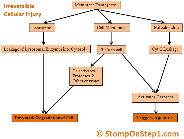
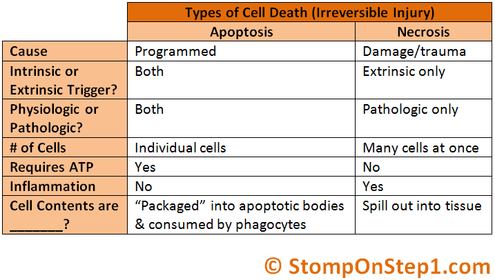
Apoptosis can be triggered by signals from within the cell (Intrinsic) or outside the cell (Extrinsic). In Intrinsic Apoptosis the cell “decides” to die because it is a normal part of physiology/development, the cell is no longer needed or the cell is too damaged to ever return to normal. Intrinsic Apoptosis signals can include decreased growth factors (genetically programmed), p53 triggers, or the release of triggers like cytochrome C as a result of cellular damage. In Extrinsic Apoptosis the cell is “told” to die by anther cell or the environment. Triggers for extrinsic apoptosis can include signals like Fas ligand on CD8 cytotoxic T cells. No matter what the original signals for apoptosis is eventually Caspace enzymes are activated. These active caspace enzymes then activate enzymes which degrade the cell.

Unlike necrosis, apoptosis can sometimes be “good” (physiologic). Problems with apoptosis arise when there is either too much or not enough.

There are various different types of necrosis that arise in different situations.
- Coagulative = Occurs in most tissues, but is most often seen in heart & kidney ischemia. Think of coagulative as the default type of necrosis that occurs if one of the specific scenarios described below is not present. Histologically cells keep their overall shape (semi-solid) but lose their nuclei resulting in a light pink area without any dark nuclei. On gross specimens it usually appears as a pale area on the organ
- Liquefactive = Present in the brain (stroke) & within abscesses (pus). Involves complete enzymatic degradation of the tissue into a liquid
- Caseous = Occurs when TB (Ghon Complex) is present or within granulomas. Appearance is a mix of coagulative & liquefactive (Caseus is the Latin term for cheese)
- Fibrinoid = Seen within the vessel wall in malignant hypertension & vasculitis. Protein from the blood leaks into the damaged vessel wall making it look pink
- Fat Necrosis = Occurs in pancreatic diseases (peripancreatic fat) or trauma to the breast. Calcium from the blood and from lysed cells mixes with the fat in the tissue to create a white soap like substance (Saponification)
- Gangrenous = Due to ischemia and/or infection (C. Perfringens). Common in the distal toes. More common in diabetics & smokers (Buerger’s Vasculitis). May require amputation
Pictures Used:
- “Apoptosis” by Emma Farmer available at http://en.wikipedia.org/wiki/File:Apoptosis.png by Public Domain
- “MI with contraction bands very high mag” by Nephron available at http://en.wikipedia.org/wiki/File:MI_with_contraction_bands_very_high_mag.jpg by Creative Commons 3.0 Attribution-Share Alike
- “Coagulative Necrosis in Kidney Tissue” by Dentl college survival kit available at http://commons.wikimedia.org/wiki/File:Coagulative_necrosis_in_kidney_tissue.jpg by Public Domin
- Derivative of “Liquefactive necrosis in brain tissue” by Daftblogger available at http://commons.wikimedia.org/wiki/File:Liquefactive_necrosis_in_brain_tissue.jpg by Public Domain
- Derivative of “Tuberculoid granuloma humpath 2” by Humpath available at http://commons.wikimedia.org/wiki/File:Tuberculoid_granuloma_humpath_2.png by Creative Commons 3.0 Attribution-Share Alike
- Derivative of “Fibrinoid necrosis in an artery” by Quizlet available at http://commons.wikimedia.org/wiki/File:Fibrinoid_necrosis_in_an_artery.jpg by Public Domain
- Derivative of “GangreneFoot” by James Heilman MD available at http://en.wikipedia.org/wiki/Gangrene by Creative Commons 3.0 Attribution-Share Alike
Now that you have finished this video you should check out the next video in the Cell Injury, Cell Death & Cancer sections which covers Free Radical Damage.
can I get some references that I can go through for this ??
The links for the pictures I used are listed at the bottom of the page.
I don’t really have any specific references for the content of the page. I’m not really citing any specific books or journal articles. This is all general material that would be covered in any pathology textbook, Step 1 review book or medical school general pathology unit of lectures. I just took that general material and organized it, explained it and presented it a bit differently than most other sources.
Does that answer your question?
Thanks!
-Brian
Thank You so much for sharing your biological Knowledge