Parasites are organisms that live in or on a host. These organisms gain some sort of survival advantage (such as gaining nutrients) while their presence is often detrimental to the host. Parasites usually don’t kill their host, but can cause disease if the parasite burden is high enough. For the exam, the most important group of parasites is Protozoa. These are microscopic unicellular eukaryotes that are motile. They move using a tail or foot like processes. Different species have a predilection for living in different parts of their human host. The most important protozoa for the USMLE Step 1 medical board exam are Malaria, Babesiosis, Toxoplasmosis, Cryptosporidium, and Giardia Lamblia. The other main group of parasites that cause disease in humans is the Helminths or worms. However, these are largely low yield material so we will just briefly cover this group towards the end of the video.
Protozoa of RBCs:
Malaria is a disease caused by the Plasmodium Protozoa parasite that is transmitted by Mosquitos. The most common species are Plasmodium Falciparum, Plasmodium Vivax, Plasmodium Ovale, and Plasmodium Malariae. Each of these has slightly different characteristics, but for the most part these differences are beyond the scope of Step 1. There is a very complex life cycle, but learning all of those details isn’t necessary for the exam. When inside a human host these parasites mainly reside inside red blood cells. Clinically, malaria presents with reoccurring cycles of spiking fevers and chills with other non-specific symptoms like headache and sweating. These “attacks” are interspersed with periods of complete remission. The paroxysmal symptomatic periods of different species of malaria occur at different frequencies, but that is beyond the scope of Step 1. In general the attacks occur every couple days or so. Symptoms occur when mature schizonts rupture erythrocytes releasing immature merozoites. Anemia may be present due to this rupture of red blood cells. The question stem almost always mentions recent travel to a place like Africa or Latin America as Malaria is not endemic to the United States. One interesting correlation is that Sickle Cell Trait offers some resistance to certain malaria species. This is why sickle cell trait and disease is much more common in area where malaria is endemic. Sickle Cell trait actually gives a survival advantage due to its antimalarial property. A peripheral blood smear will show enlarged RBCs with numerous small parasite “dots” on Giemsa stain. Being able to identify the different specific species of plasmodium based on histology is not necessary for the exam.


Antimalarials are a class of medication that can be used prophylactically to prevent malaria, used to treat identified/suspected malaria, or used to periodically treat populations in endemic areas. Most of the antimalarials have “Quine” in the name which makes them easy to pick out of a list. The most commonly used antimalarials are Chloroquine, Hydroxychloroquine, Mefloquine, & Primaquine. Quinine is primarily used for severe cases of malaria. Doxycyline also has some action against malaria and is most often used for prophylaxis. The mechanisms of these drugs is beyond the scope of the USMLE Step 1 exam.
Resistance to antimalarial medications is common and a huge public health concern. Different areas of the world have different resistance patterns which helps dictate which drugs should be used as first line treatments, but knowing those details is beyond the scope of Step 1. Combination therapy is often used now to prevent resistance and recurrence. For example, combining chloroquine or hydroxychloroquine with primaquine is a common combination treatment. Monotherapy is usually sufficient for chemoprophylaxis.
There are also non-pharmacologic preventative measures. Spraying insecticides, using personal protective clothing/mosquito repellent, draining areas of standing water (to destroy mosquito habitats), and using bed nets can decrease patients exposure to mosquitos.
Babesia Microti is a protozoa infection of the red blood cells similar to malaria. It is usually asymptomatic, but in some patients babesiosis can present with similar symptoms to malaria. It is transmitted by the Ixodes Tick that is also the vector for Lyme disease. Like Lyme disease, Babesia is largely found in the northeast US.
GI Protozoa
The gastrointestinal protozoa include Cryptosporidium, Giardia Lamblia & Entamoeba Histolytica. All 3 are primarily spread via ingestion of cysts in contaminated water (AKA fecal-oral transmission) and all 3 present with diarrhea. Cryptosporidium presents as watery diarrhea primarily in AIDs patients. Oocysts may be found in the stool on acid fast stain and only symptomatic treatment is needed. Giardia Lamblia presents with bloating, cramping, flatulence and fatty floating stools. Recent camping or hiking is often mentioned in the question stem. On stool histology giardia can be seen as a symmetric pear shaped trophozoite. Metronidazole is the treatment of choice. Entamoeba histolytica presents as bloody diarrhea and intestinal ulcerations. Histologic examination of the stool shows trophozoites which have ingested red blood cells. Metronidazole is the treatment of choice.


Metronidazole is an antimicrobial medication that has antiprotazoal activity against Trichomonas, Giardia Lamblia & Entamoeba Histolytica. It is also frequently used for C. Dif, Bacterial Vaginosis, and H. Pylori. A common side effect is inhibition of alcohol metabolism. This “Disulfiram-like” reaction leads to an exaggerated effect even when a small amount of alcohol is consumed. You will remember from our earlier video on alcohol metabolism that Disulfiram is a medication used to treat alcoholism as it gives them a really bad hang over if they drink any alcohol. Despite the “Azole” ending to the name metronidazole is not used as an antifungal medication like many other Azoles.
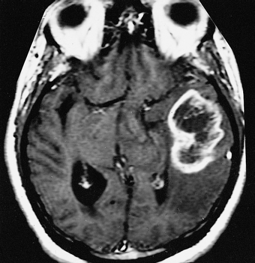
Toxoplasmosis (Toxoplasma Gondii) is a parasite that primarily causes brain abscesses/cysts in HIV patients with a CD4 T Cell count <200. In the united states this protozoa usually infects a human host after they eat undercooked pork or are exposed to cat feces. On MRI or CT scan the lesion(s) presents as a “ring enhancing” (low density circular area surrounded by a white/high density rim). Infections are usually asymptomatic and then can become reactivated when the patient becomes immunocompromised. The symptoms depends on where the lesion is. Toxoplasmosis can present as a headache with a wide variety of neurological deficits, but seizures are most common.

Pinworm (AKA Enterobius Vermiculus) is a type of roundworm/nematode. Infection occurs through the ingestion of eggs in food. It is the most common helminth infection in the US and is primarily seen in children. Patients are asymptomatic except for severe perianal itching. This itching is caused when the worms, that live in the intestine, migrate during the night and deposit their eggs outside of the anus. Diagnosis is made by “Cellophane Tape Test” (AKA scotch tape test). Here a piece of clear adhesive tape is applied to the perianal area and then examined microscopically to look for eggs or adult worms.
Mebendazole or Albendazole are the first line treatment for most worm infections. They are both broad spectrum anti-helminth that inhibits microtubule formation and decrease nutrient uptake by the worm. Dispite the “Azole” ending to the word, these medications are not in the antifungal azole class. A mnemonic to remember these medications is that WORMs are BENDy.
Pictures Used:
- “Malaria lifecycle-CDC” by CDC available at https://commons.wikimedia.org/wiki/File:Malaria_lifecycle-CDC.gif via Public Domain
- Derivative of “Malaria in Peripheral Blood (6289093848)” by Ed Uthman available at https://commons.wikimedia.org/wiki/File:Malaria_in_Peripheral_Blood_(6289093848).jpg via Creative Commons 2.0 Generic License
- Derivative of “Micrograph depicts a number of ring form plasmodium falciparum trophozoites royalty free photo” by Steven Glenn available at http://www.public-domain-image.com/free-images/science/microscopy-images/malaria-plasmodium/micrograph-depicts-a-number-of-ring-form-plasmodium-falciparum-trophozoites/attachment/micrograph-depicts-a-number-of-ring-form-plasmodium-falciparum-trophozoites via Public Domain
- “Cryptosporidium DPDxCrypto oo AF” by CDC available at https://commons.wikimedia.org/wiki/File:Cryptosporidium_DPDxCrypto_oo_AF.JPG via Public Domain
- “Giardia lamblia cytology closeup” by Jerad M Gardner available at https://commons.wikimedia.org/wiki/File:Giardia_lamblia_cytology_closeup.jpg via Creative Commons 3.0 Unported Attribution-Share Alike License
- “Trophozoites of Entamoeba histolytica with ingested erythrocytes” by CDC available at https://commons.wikimedia.org/wiki/File:Trophozoites_of_Entamoeba_histolytica_with_ingested_erythrocytes.JPG via Public Domain
- “Brain Abscess at MRI (T1 + contrast)” by Aimun AB Jamjoom available at via https://commons.wikimedia.org/wiki/File:Brain_Abscess_at_MRI_(T1_%2B_contrast)_–_showing_a_small_ring-enhancing_lesion_with_mild_surrounding_edema_adjacent_to_the_ventricular_catheter_and_ventricular_dilatation..jpg Creative Commons 2.5 Generic License
- “AFIP-00405558-Glioblastoma-Radiology” by Armed Forces Institute of Pathology available at https://commons.wikimedia.org/wiki/File:AFIP-00405558-Glioblastoma-Radiology.jpg via Public Domain
- “Tape dispenser” by Donmike10 available at https://commons.wikimedia.org/wiki/File:Tape_dispenser.JPG via Public Domain